
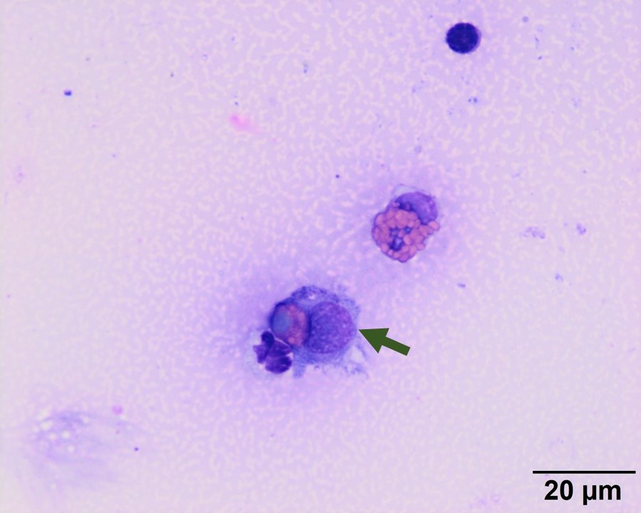
A macrophage/synoviocyte (arrow) is phagocytizing an eosinophil. Note how pale and smooth the nuclear chromatin of the phagocytized eosinophil appears to be. This finding is part of the normal enzymatic cellular degradation after a phagocyte engulfs an extravasated leukocyte (Wright’s stain, 100x objective).