
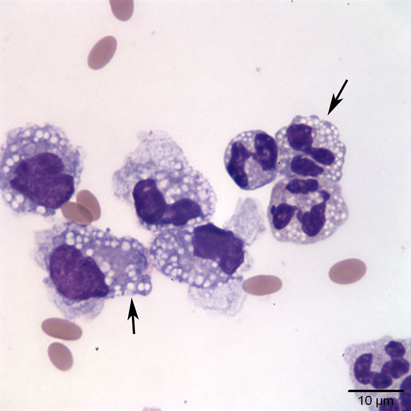
Figure 3a. Peritoneal fluid from an alpaca with outflow obstruction highlighting discrete, margined cytoplasmic vacuoles in the inflammatory cells, compatible with lipid (Wright’s stain, 100x objective)
Figure 3a. Peritoneal fluid from an alpaca with outflow obstruction highlighting discrete, margined cytoplasmic vacuoles in the inflammatory cells, compatible with lipid (Wright’s stain, 100x objective)
eClinpath helped 1.2 million visitors last year from 220 countries find important information on animal health. If you enjoy the site, please support our mission and consider a small gift to help us keep pace with its rapid growth. You can donate securely via PayPal or credit card. Thank you!
![]()