
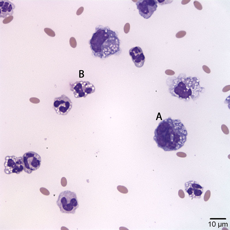
Figure 2a. Peritoneal fluid from an alpaca with outflow obstruction highlighting a macrophage (A) and a non-degenerate neutrophil (B). Note the discrete, margined vacuoles in the cytoplasm of the cells, compatible with lipid. (Wright’s stain, 50x objective)