
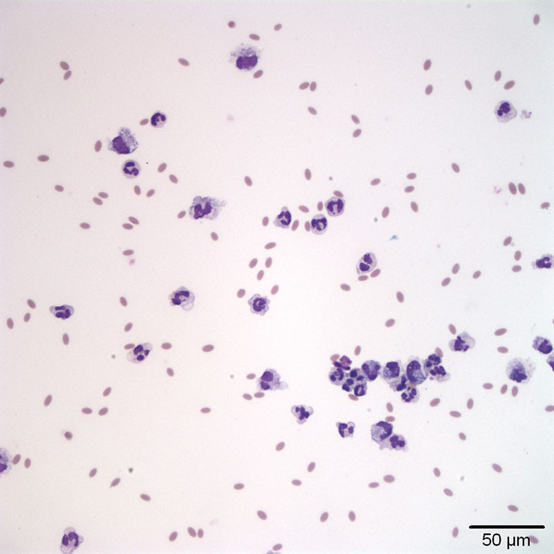
Figure 1a. Peritoneal fluid from an alpaca with outflow obstruction. There is a mixture of inflammatory cells, predominated by non-degenerate neutrophils with fewer macrophages in a background of a small amount of blood (Wright’s stain, 20x objective).