
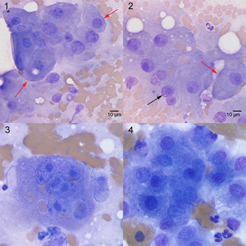
Figure: In image 1, the cells demonstrate moderate to marked anisokaryosis and several have nuclei that contain more than one nucleolus. There is also a tendency for the cells to dissociate from the main cluster (individualization; red arrows). In image 2, hepatocytes have large nucleoli (macronucleoli, the same size or larger than the red blood cells in the background, one example is shown with the black arrow), multiple nucleoli, or are becoming individualized (red arrow). A multinucleated giant cell (with macronucleoli or multiple nucleoli of variable size) is seen in image 3. In contrast, in the aspirate from the liver of a dog with lymphoma (image 4), the hepatocytes display no cytologic abnormalities. They are relatively uniform in appearance with prominent pink granulation to their cytoplasm. They have central to paracentral round nuclei and contain single small nucleoli (not prominent in the image). Neoplastic lymphocytes were not identified in the aspirate.