
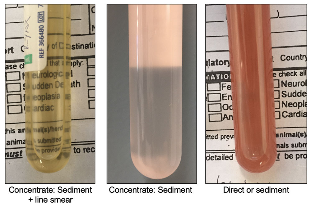
The fluid on the left is light yellow and clear; the writing on the page can be read easily through the fluid. Such samples are of low cellularity and require concentration techniques to optimize smear preparation and cytologic evaluation, ideally cytocentrifugation. In the absence of a cytocentifuge, a sediment can be prepared from the sample with a regular centrifuge and a line smear prepared from the resuspended pellet to maximally “concentrate” the cells. A line smear is similar to a wedge smear used for blood smear preparation, but instead of creating a feathered edge, the spreader slide is lifted up before the fluid is fully smeared to create a “line” of fluid. The middle tube is light pink and slightly cloudy. The latter appearance reflects either cells or lipid, with the pink tinge supporting the presence of red blood cells. Optimal smear preparation would involve concentration via centrifugation, although smears of unconcentrated fluid (direct smears) will be more cellular than that from the tube on the left. Centrifugation would also help discriminate between lipid (would not sediment) and cells (a sediment would form) or reveal the presence of both (cloudy supernatant with or without a fat layer plus a cell pellet). If lipid is present, triglyceride concentrations on the fluid could be measured to help support leakage of chyle as a component of the effusion. The tube on the right is light to medium red and opaque, preventing visualization of the text on the page behind the tube. The color indicates the presence of intact red blood cells, which will sediment on centrifugation. Nucleated cellularity (e.g. white blood cells) may vary from low to high. For this type of sample, a direct smear of unconcentrated fluid and a sediment smear of a resuspended pellet of centrifuged fluid would be optimal smear types.