
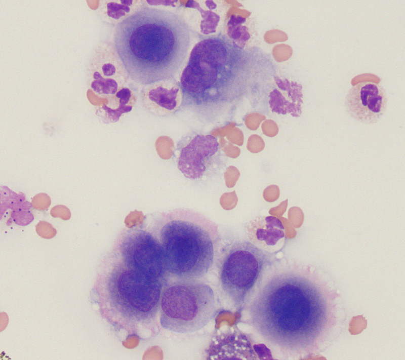
Numerous mesothelial cells are seen in this pleural fluid from a dog with a transudative effusion (with concurrent diapedesis of red blood cells or hemorrhage). The mesothelial cells have central round nuclei with a moderate amount of light purple cytoplasm and a “corona” or “fringe” to the cytoplasmic borders. Binucleated and multinucleated forms may be seen if the cells are reactive, along with clusters of mesothelial cells with deeper blue cytoplasm. The mesothelial cells in this fluid resemble those seen in many non-neoplastic effusions and essentially lack cytologic criteria of malignancy, despite this fluid being from a dog with documented mesothelioma (based on tissue invasion in surgical biopsies of tissues). In this dog, they were likely neoplastic despite resembling normal or reactive counterparts.