
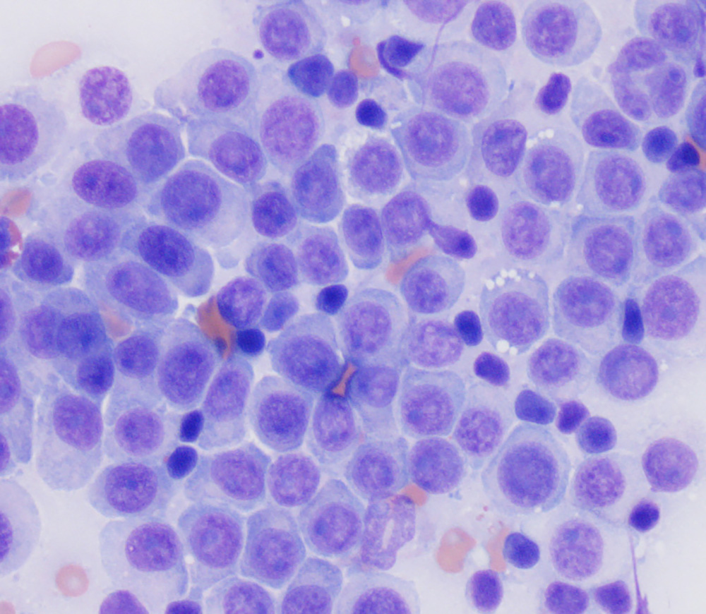
This was an aspirate of a flank transmissable venereal tumor in a dog. The cells are round with quite high nuclear to cytoplasmic ratios and distinct cell borders. The nuclear chromatin is clumped and mitotic figures were evident (not shown). There is a concurrent infiltrate of small lymphocytes. Only low numbers of cells have the characteristic discrete margined cytoplasmic vacuoles (Wright’s stain, 50x objective).