
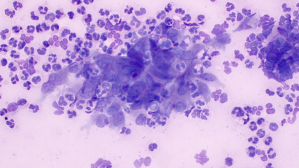
Corneal scraping from a horse with an ulcer” There is marked neutrophilic inflammation and neutrophils are closely intercalated with the corneal epithelial cells. The atypical features of the cells are higher nuclear to cytoplasmic ratios and deeper blue cytoplasm than normal. They also display mild anisokaryosis and have irregular nuclear and cellular shapes, with several cells being more spindled. These features were attributed to a reactive response secondary to the inflammation versus a tumor, such as a squamous cell carcinoma (they were not keratinizing). Some pathologists may use the term dysplasia instead of atypia for these cellular features (Wright’s stain, 50x objective).