
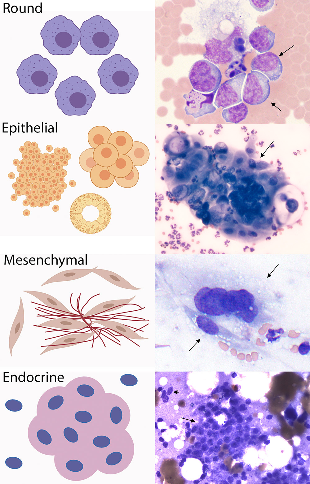
The left panel shows a cartoon, whereas the right panel shows an example image from a cytologic smear of different tumor types (in this case, all images are from a horse). The different tumors are distinguished cytologically as: a) Round cell : Exfoliate individually as round cells and usually of hematopoietic origin, e.g. lymphoma (arrowed cells), mast cell tumor, histiocytic tumors, transmissable venereal tumors; b) Epithelial cells: Exfoliate as clusters, sheets or, rarely, acini but can be seen as individual cells. Cells are often round to polygonal with distinct boundaries, e.g. squamous cell carcinoma (arrow), trichoblastoma, gastric carcinoma, perianal gland adenoma; c) Mesenchymal: Exfoliate as individual cells with tapering boundaries and indistinct borders. Can be seen in aggregates, potentially intercalated with strands of matrix, e.g. hemangiosarcoma (no matrix, arrows – the upper cell is multinucleated), soft tissue sarcoma, melanoma, fibrosarcoma; d) Endocrine/neuroendocrine: Exfoliate as packets or nests (arrow) and rupture easily leaving bare nuclei (arrowhead) in the background of ruptured cytoplasm, e.g. thyroid adenoma (shown) or carcinoma, apocrine anal sac adenocarcinoma, gastrinoma (modified Wright’s stain)