
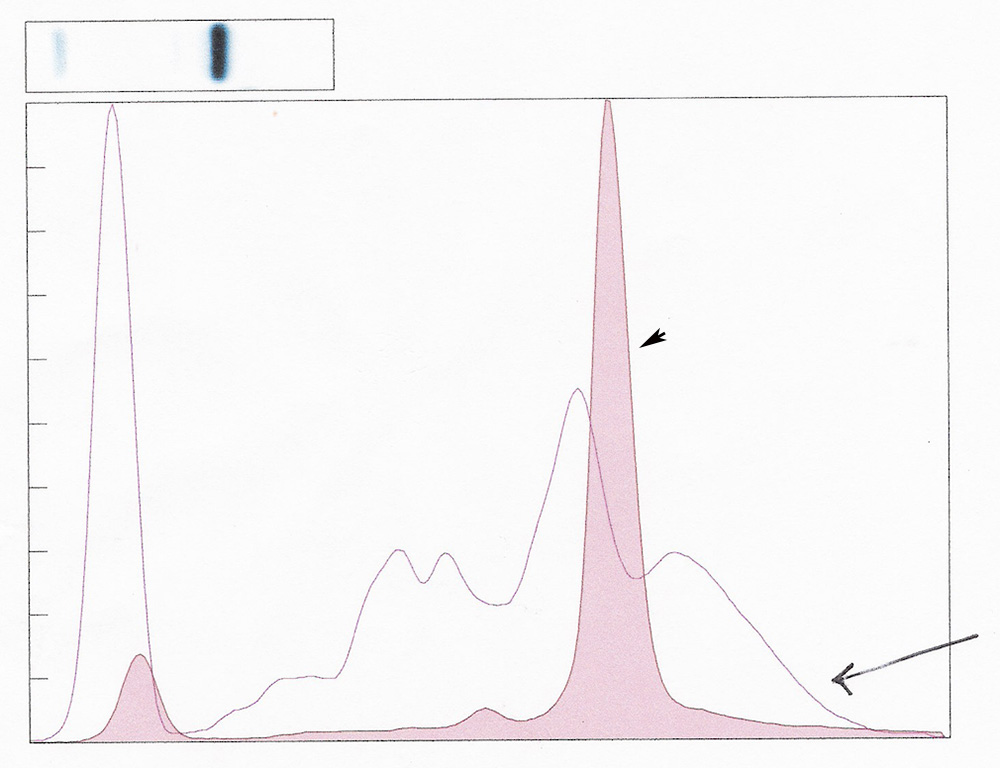
A red blood cell lysate was made to generate hemoglobin, which was run on the agarose gel. This was overlaid with a serum electrophoretogram from a dog, in which the sample was hemolyzed. Note how the hemoglobin in the red blood cell lysate (arrow, pink solid curve) aligns with the beta-2 spike in the dog’s serum electrophoretogram (open gray curve, arrow).