
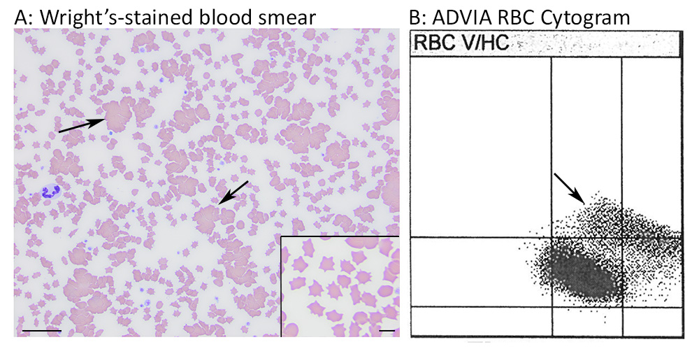
A: Red blood cell agglutinates (arrows) are present in the modified Wright’s-stained blood smear (bar = 25 um). The higher magnification inset (scale bar = 5 um) shows that the red blood cells lack features that may be seen with an IMHA, including spherocytes (hard to conclusively identify in cats) and polychromasia (expected in those patients that are regenerating). Indeed there were moderate to many echinocytes and low numbers of acanthocytes and echino-elliptocytes. These RBC changes are not specific but could be due to the chronic kidney disease in the cat. B: The ADVIA 2120i RBC cytogram or “tic-tac-toe” plot shows a separate population of RBCs from the normocytic normochromic cluster (in the center box) that is spanning the macrocytic normochromic to the macrocytic/normocytic hyperchromic boxes (arrow). The separate cluster is compatible with red blood cell agglutinates.