
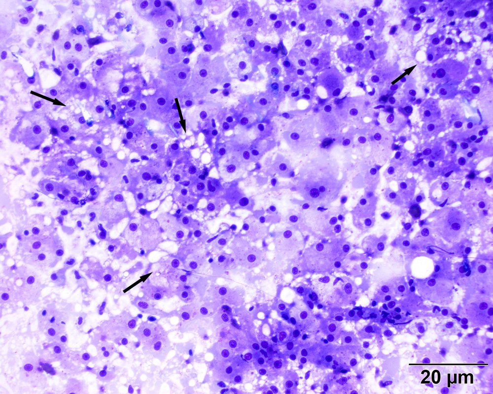
The large neoplastic cells are round to polygonal and seen as a cohesive-like sheet. Several cells contain clear discrete-margined vacuoles, most consistent with lipid (arrows). Lipid is also present in the background, likely from r ruptured cells (Diff-quik stain, 20x objective)