
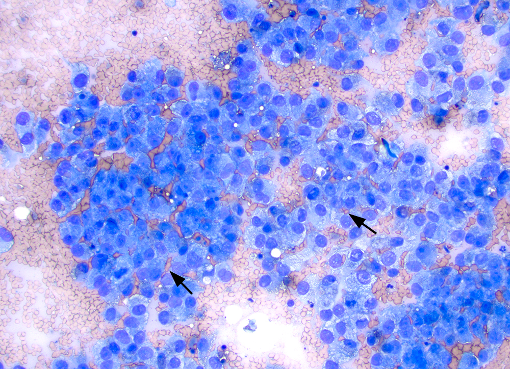
From low power, the aspirate contained aggregates and sheet-like arrangements (depicted here) of medium to large mostly round to oval cells. Although the cells appear cohesive, red blood cells are intercalated between the tumor cells (arrows as examples), arguing against cohesion and an epithelial tumor. A mitotic figure is present near to the arrowed red blood cells on the left. There is a hint of cellular necrosis in the background and a fragment of a hemosiderophage (center top left of image). 20x objective, modified Wright stain.