
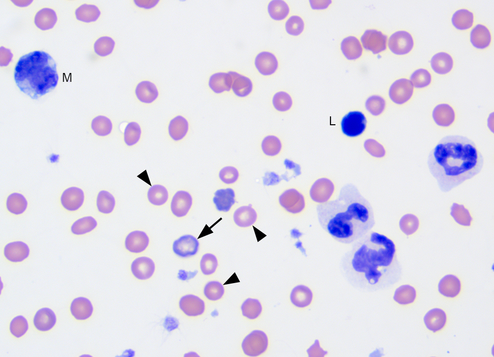
A higher power image of the blood smear shows a hypochromic polychromatophil (arrow) and mature red blood cells (arrowheads). There are three neutrophils, two of which have small Dohle bodies (can be normal in cat blood, but neutrophils were still judged to be mildly toxic). The neutrophils are showing storage-associated changes, including lightening and swelling of the nuclear chromatin (resembling band forms; all of the depicted cells would have been counted as mature segmented neutrophils), clear discrete margined cytoplasmic vacuoles, and polarization of neutrophil granules (the sample was collected the night before and freshly prepared smears were not provided with the blood in the EDTA-anticoagulant tube). Regardless, band neutrophils were identified in the blood smear, indicating an inflammatory leukogram. The other two leukocytes are a monocyte (M) and small lymphocyte (L). 100x objective, modified Wright stain