
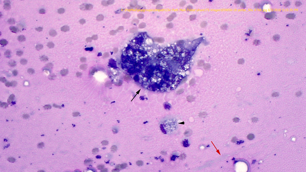
Higher power image of the previous figure, illustrating the multinucleated macrophage, which contains numerous lipid droplets and erythrocytes in the cytoplasm (black arrow) and linear strands of light blue mucus (red arrow). An adjacent mononuclear macrophage also contains cytoplasmic lipid droplets (50x objective, Wright’s stain).