
Aspirates from an abdominal and cutaneous mass in a dog
Case information
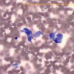
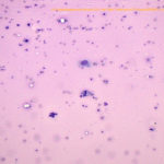
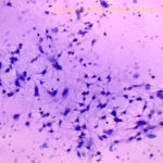
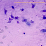
A neutered 8 year old Staffordshire Terrier mixed breed dog presented for re-evaluation of skin masses. Two subcutaneous (presumably ultraviolet light-induced) hemangiosarcomas had been surgically excised in the past. On examination, the dog had multiple red cutaneous lesions on the medial aspect of both hindlimbs and similar small lesions on the caudal abdomen. In addition, the dog had a subcutaneous mass on the right lateral distal hindlimb. Imaging for tumor staging revealed an abdominal mass cranial to the bladder. Aspirates were taken from two of the red cutaneous lesions (one on the right medial hind leg, one on the left medial hind leg), the subcutaneous mass on the right lateral distal hind leg and the abdominal mass. Examine the images of the smears from the right lateral distal hind leg and abdominal mass and answer the questions below.
- What types of cells are found in the smears from each mass?
- What is your cytologic diagnosis for each site?
- Do you think the abdominal mass is related to the skin mass?
|
|
|
|
|
Answers on next page