
Aspirate from a subcutaneous mandibular mass in a cat
Case information
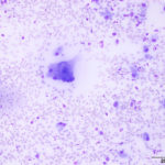
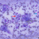
An 18 year old male neutered Domestic Shorthair cat presented to the Cornell University Hospital for Animals (CUHA) Emergency Service for evaluation of a subcutaneous left mandibular mass and inappetence. The mass had been previously incised by the primary veterinarian and had yielded purulent material. At that time, treatment with doxycycline was started, however the mass continued to increase in size. At the time of presentation to CUHA, the mass was approximately 2 x 2 x 0.5 cm, not painful on palpation, and not freely movable. Fine needle aspirates were taken from the mass for cytologic examination. Examine the representative images of the smears and answer the questions below:
- What are the cells marked by the red and blue arrows in Figure 2?
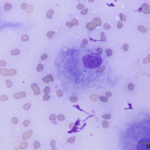
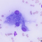
- What are your top differential diagnoses for the structures within the cells in Figures 3 and 4?
- What additional testing would you recommend?
 |
 |
 |
 |
Answers on next page
Pages: 1 2