
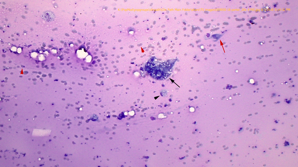
The aspirate consisted largely of light pink mucinous material, moderate amounts of blood, low to moderate macrophages and numerous spindle cells with some abdominal fat. The mucinous material delineated erythrocytes and leukocytes into streams (called windrowing, red arrowheads). Spindle cells were individualized (red arrow) and many were caught up within mucinous matrix (Figure 3). They displayed mild to moderate anisocytosis and anisokaryosis. Macrophages (black arrowhead), which contained phagocytized lipid and/or erythrocytes in their cytoplasm, were seen throughout the smear, with some giant multinucleated versions (black arrow) (Wright’s stain, 20x objective)