
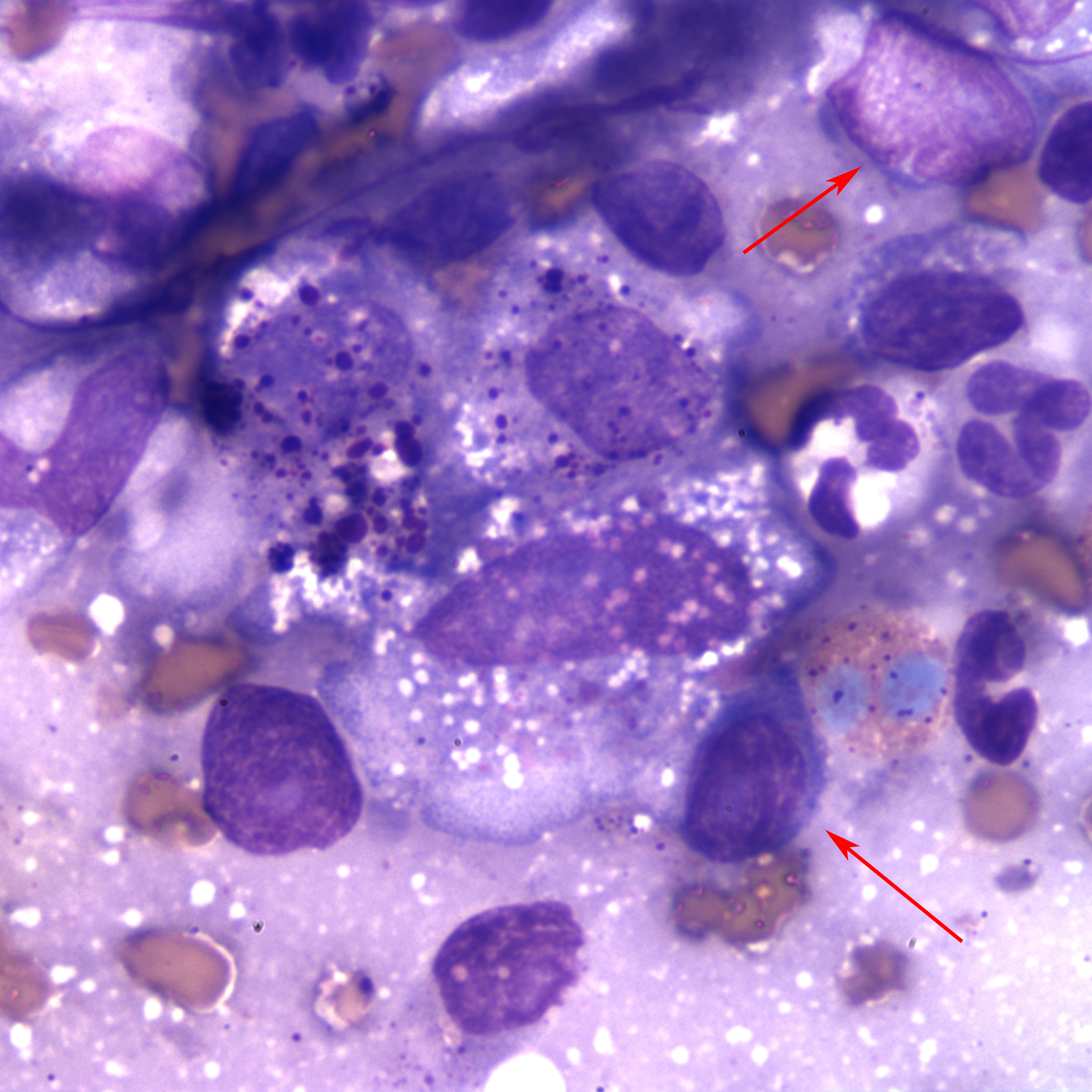
Figure 4. Liver mass in a cat (Wrights stain, 100x). Three large, vacuolated macrophages are present in the center of the image, containing variable numbers of fine to globular magenta cytoplasmic granules. The red arrows also point out more examples of atypical (neoplastic) lymphocytes in this case.