
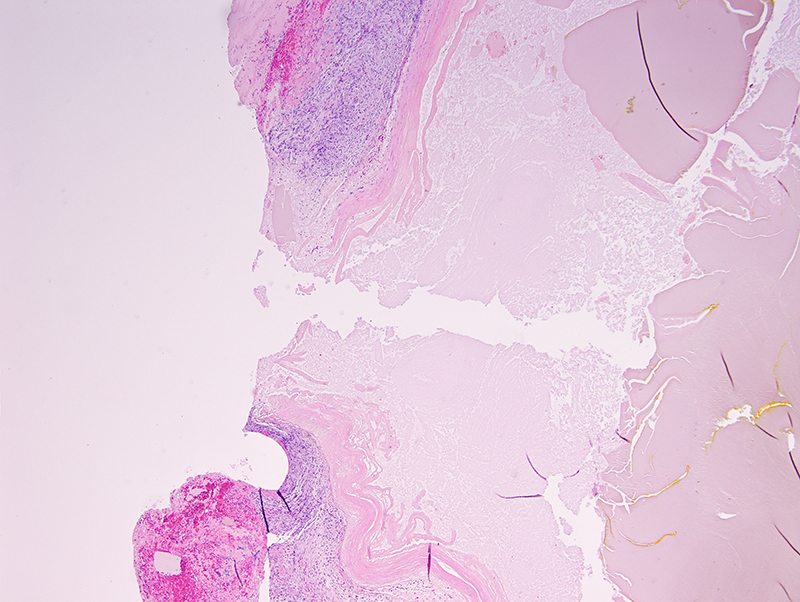
Histologic section of this patient’s gallbladder mucocele showing the full thickness defect in the gallbladder wall. There is a fibrinous and hemorrhagic clot on the outer wall of the gallbladder (lower left) (H&E, 4x). Photo courtesy of Dr. Mason Jager.