
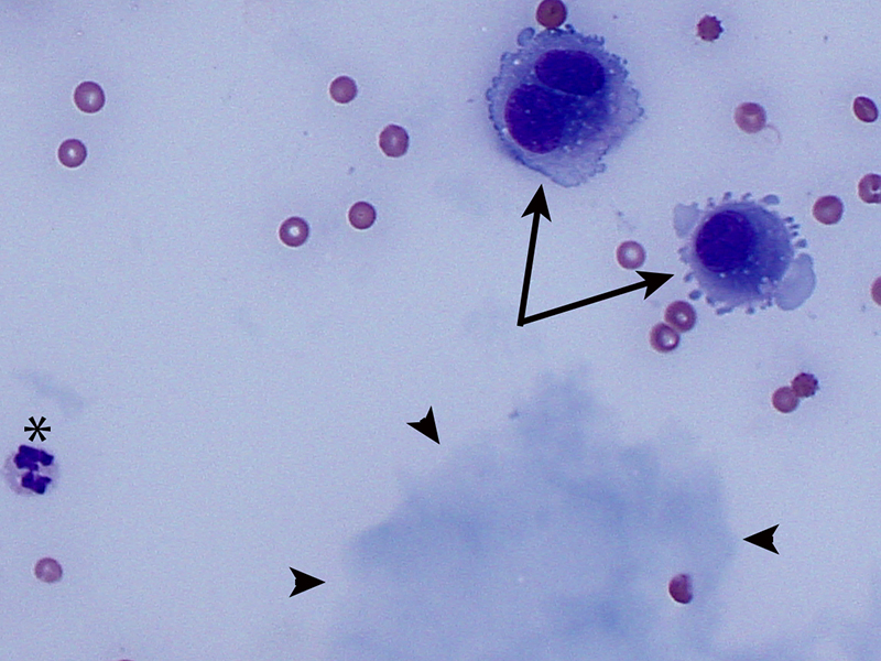
A few reactive mesothelial cells (arrows) were present in the sample. These are commonly seen in smears of effusions from dogs. The reactive features include the binucleation and deeply basophilic cytoplasm. A lake of mucin/”white bile” is visible (arrowheads). A neutrophil is also present (asterisk). (Direct smear, Wright’s stain, 50x)