
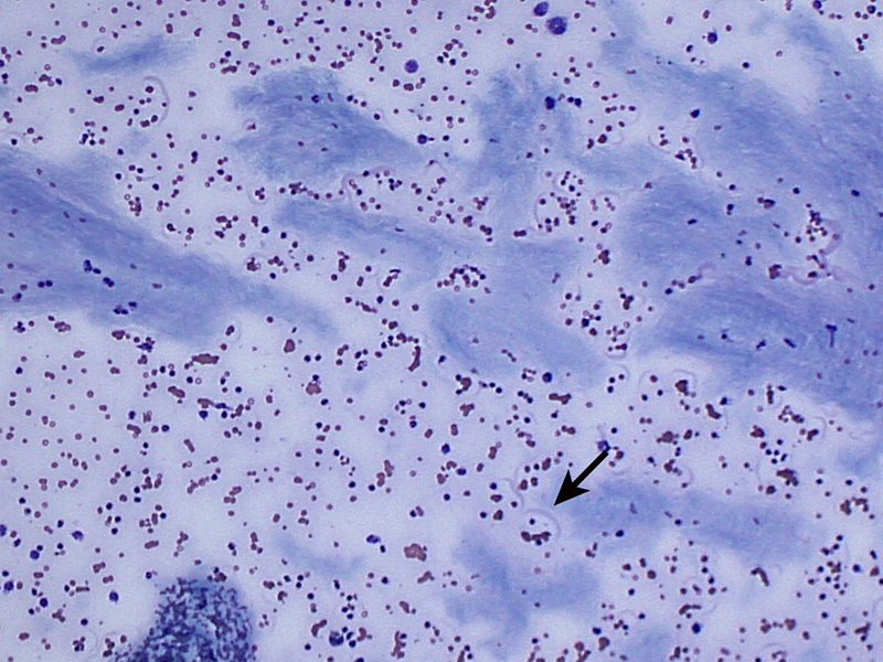
Additional low power view showing abundant amounts of the light blue amorphous extracellular material consistent with mucin or “white bile.” The arrow is pointing to a protein crescent evident on the smear supporting an increased protein content of the peritoneal fluid (Direct smear, Wright’s stain, 10x).