
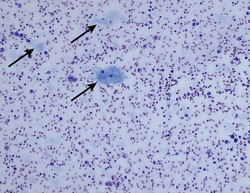
Representative low power view showing the extracellular light blue amorphous material consistent with mucin or “white bile,” indicated by the arrows. (Direct smear, Wright’s stain, 10x)
Representative low power view showing the extracellular light blue amorphous material consistent with mucin or “white bile,” indicated by the arrows. (Direct smear, Wright’s stain, 10x)
eClinpath helped 1.2 million visitors last year from 220 countries find important information on animal health. If you enjoy the site, please support our mission and consider a small gift to help us keep pace with its rapid growth. You can donate securely via PayPal or credit card. Thank you!
![]()