
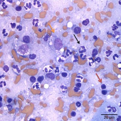
Figure 3b: The inflammatory cells are mostly mildly degenerate neutrophils (black arrow) with fewer macrophages (yellow arrow), small lymphocytes (red arrow), and plasma cells (not shown) (Wright’s stain, 500x magnification).
Figure 3b: The inflammatory cells are mostly mildly degenerate neutrophils (black arrow) with fewer macrophages (yellow arrow), small lymphocytes (red arrow), and plasma cells (not shown) (Wright’s stain, 500x magnification).
eClinpath helped 1.2 million visitors last year from 220 countries find important information on animal health. If you enjoy the site, please support our mission and consider a small gift to help us keep pace with its rapid growth. You can donate securely via PayPal or credit card. Thank you!
![]()