
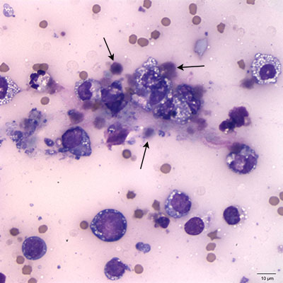
Figure 7: Pericardial fluid, direct smear. Lymphoma cells with necrotic cellular debris. The arrows are pointing to the areas of necrosis (500x, Wright’s stain)
Figure 7: Pericardial fluid, direct smear. Lymphoma cells with necrotic cellular debris. The arrows are pointing to the areas of necrosis (500x, Wright’s stain)
eClinpath helped 1.2 million visitors last year from 220 countries find important information on animal health. If you enjoy the site, please support our mission and consider a small gift to help us keep pace with its rapid growth. You can donate securely via PayPal or credit card. Thank you!
![]()