
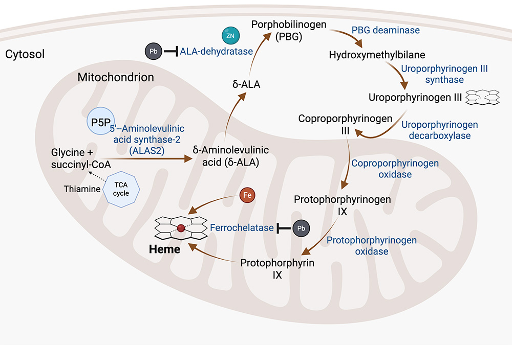
Heme synthesis occurs in the mitochondria and cytosol of developing nucleated erythroid progenitors. The first step in the pathway is the formation of delta-aminolevulinic acid (d-ALA) from glycine and succinyl-CoA by the enzyme 5’-aminolevulinic acid synthase (ALAS) in the mitochondrion. There are two isoforms of ALAS, with isoform ALAS2 being found in erythrocytes. Pyridoxal-5’-phosphate (P5P), the biologically active metabolite of vitamin B6, functions as an essential cofactor for ALAS2, with this first step of the heme synthesis pathway being the rate-limiting step. In the second step, d-ALA moves into the cytosol and two molecules are condensed by the enzyme ALA dehydratase (ALAD; also known as porphobilinogen synthetase) to form porphobilinogen (PBG). ALA dehydratase requires zinc (Zn) as a structural cofactor and catalyst. In steps three and four, PBG deaminase condenses four PBG molecules to the linear four-pyrrole (tetrapyrrole) unstable molecule, hydroxymethylbilane, and then uroporphyrinogen III synthase changes hydroxymethylbilane to a cyclical form, also called uroporphyrinogen III, in the cytosol. In the fifth step, uroporphyrinogen III is converted to coproporphyrinogen III by the enzyme, uroporphyrinogen decarboxylase, which removes four carboxyl groups from the acetic side chains, changing them to methyl side chains. Coproporphyrinogen III then moves into the mitochondria. In step 6, protoroporphyrinogen oxidase decarboxylates two propionate to vinyl side chains on the molecule, creating protoporphyrinogen IX, which is a reduced non-aromatic tetrapyrrole. Protoporphyrinogen IX is converted into protophoryrin IX, an oxidized aromatic tetrapyrrole, by the enzyme protoporphyrinogen oxidase, which removes six hydrogen atoms. In the final step of heme synthesis, the enzyme ferrochelatase combines ferrous (Fe2+) iron into the porphyrin or tetrapyrrole ring to create heme. Lead (Pb) inhibits the enzyme ALAD by displacing zinc from its active site. It also inhibits ferrochelatase, preventing iron from being incorporated into porphyrin ring. Lack of vitamin B6 markedly slows heme synthesis, since ALAS2 catalyzes the rate-limiting step of the heme pathway. Both lead toxicity and vitamin B6 deficiency result in accumulation of iron in RBCs, usually within mitochondria, which manifest as siderotic granules in Romanowsky-stained smears. Anucleated and nucleated RBCs that contain these granules are called siderocytes and sideroblasts, respectively. Ring sideroblasts have at least 5 siderotic granules surrounding and encompassing more than one third of the nucleus. Thiamine indirectly influences heme synthesis by serving as a cofactor for alpha-ketoglutarate dehydrogenase, a multi-enzyme complex in the tricarboxylic acid (TCA) cycle, which generates succinyl-CoA, the building block of d-ALA. Created in https://BioRender.com