
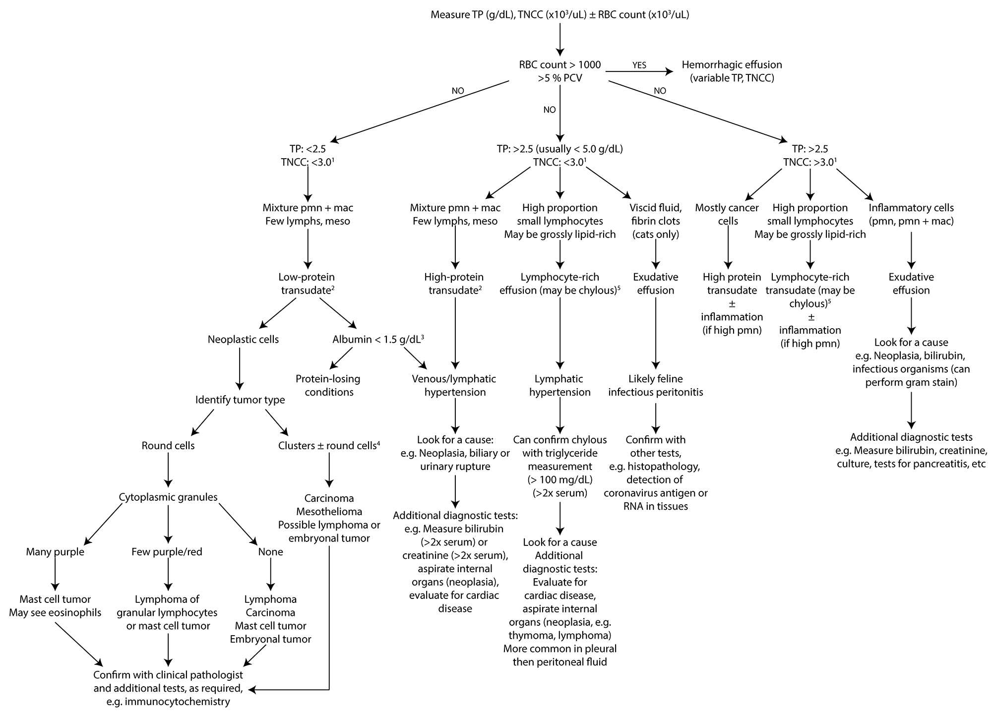
Interpretation of peritoneal and pleural fluid in dogs and cats is based on a combination of cytologic assessment of smears, total protein (by refractometer) results and total nucleated cell counts (TNCC). Red blood cell (RBC) counts can be helpful, if provided. There are three main mechanisms for an effusion: transudation (alterations in hydrodynamic forces), exudation and vessel/viscus rupture or leakage.
Note that the RBC count may be high from blood contamination (expect to see platelets in smears, if sufficient blood, sample will clot in a red top tube) or hemorrhage or erythrodiapedesis (expect to see no or few platelets, erythrophages, hemosiderophages and hematoidin crystals [in a hypoxic environment], if sufficient blood, sample will not clot in a red top tube). Hemorrhage can complicate effusions from other causes (usually exudative) and cannot be differentiated from erythrodiapedesis (due to transudation).
1 Total nucleated cell counts do not equal leukocytes, because the count also include neoplastic or mesothelial cells and, in cases of enterocentesis, bacterial clumps and food debris. Thus, counts should never be used alone to classify effusions and cytologic assessment should always be performed. Note that counts between 3,000-5000/uL are considered equivocal
2 Classification of fluid as a low or high protein transudate is mechanistically informative for peritoneal fluid, with leakage of low protein intestinal lymph (lymphangiectasia, presinusoidal portal hypertension) resulting in a low protein transudate and leakage of high protein hepatic lymph (sinusoidal or post-sinusoidal portal hypertension) in a high protein transudate.
3 Low albumin (<1.5 g/dL) can be seen in cats and dogs with severe protein-losing nephropathy or enteropathy and, less frequently, severe hepatocellular insufficiency. Usually, an effusion in an animal with mild to moderate low albumin concentrations is caused by concurrent venous or lymphatic hypertension or obstruction.
4 Clusters and individual large round cells in a fluid can be mesothelial cells (reactive or neoplastic) or from round cell tumors (e.g. lymphoma). We have also seen primitive embryonal tumors (nephroblastoma) exfoliate as individual cells with small adherent clusters, mimicking a carcinoma.
5 Due to high chylomicron content which scatter light, the total protein concentration by refractometer will be inaccurate. Note that total protein concentrations can be greater than or less than 2.5 g/dL in non-chylous lymphocyte-rich effusions, depending on the cause of the effusion.
TP: Total protein by refractometer, TNCC: Total nucleated count, RBC: Red blood cell count, PCV: Packed cell volume, PMN: Neutrophils, Mac: Macrophages, Meso: Mesothelial cells.