
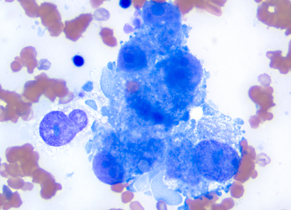
The tumor cells have distinct spindled shapes, supporting a mesenchymal tumor. The red blood cell appears phagocytized, although it could also be within a lumen between the cells (arrow). Note the anisokaryosis and anisocytosis of the tumor cells and prominent large nucleoli (1-2 per cell) 100x objective, modified Wright stain.