
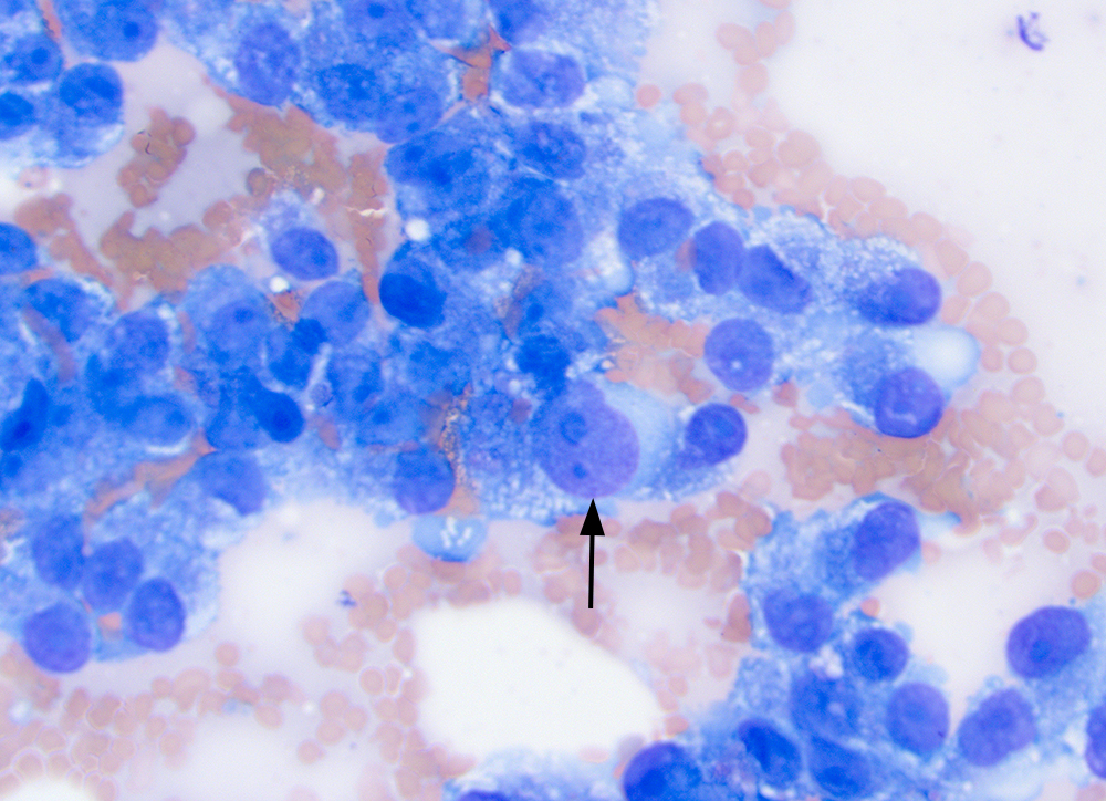
The individual tumor cells display moderate to marked anisocytosis and anisokaryosis. Red blood cells are intercalated between the individual tumor cells, a feature that argues against an epithelial tumor. We do see this cytologic pattern with hemangiosarcomas, particularly epithelioid variants, which can mimic epithelial tumors. The arrowed cell has a distinct spindled appearance and two nucleoli, one of which is quite large (macronucleolus) and abnormally shaped. Note the microvesiculation in the tumor cells and medium to dark blue cytoplasm. 50x objective, modified Wright stain.