
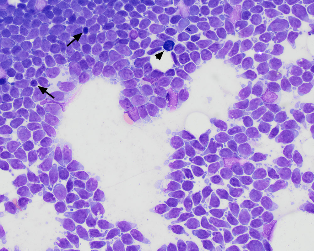
A modified Wright-stained smear shows the neoplastic cells, which dominated in aspirates of both the prescapular and popliteal lymph nodes. Nucleoli and the fine chromatin are more apparent in this stain as are cytoplasmic vacuoles. Note the dark and clumped of residual small lymphocytes (arrows). A single plasma cell is present (arrowhead) (50x objective)