
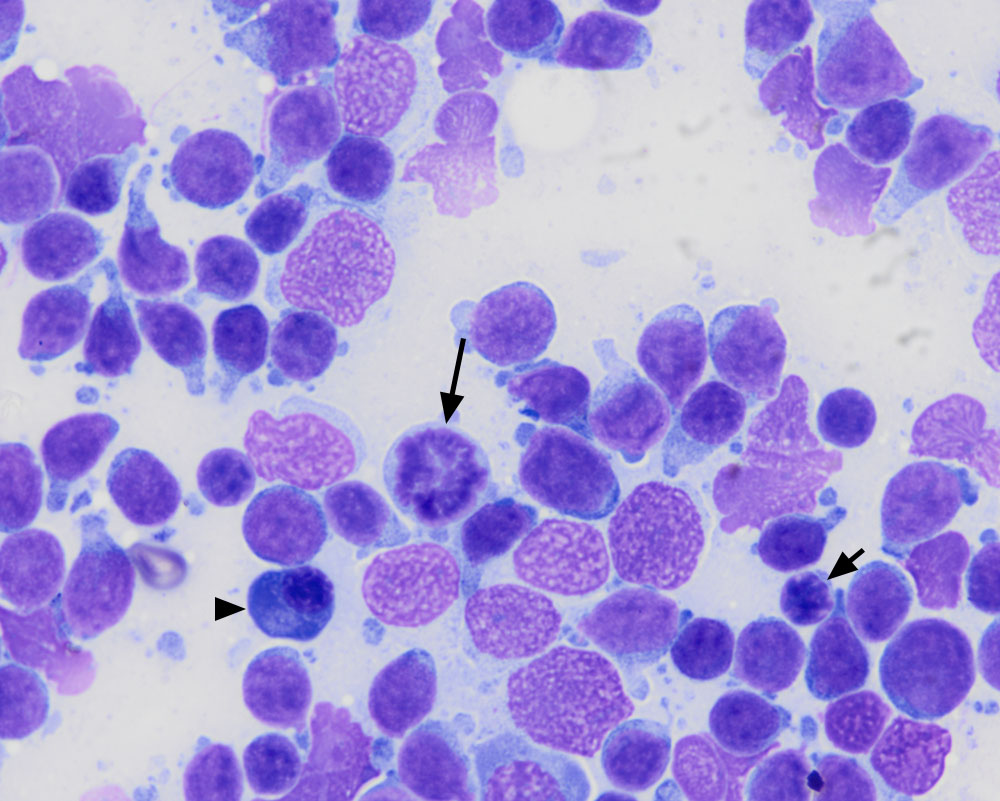
This high magnification shows the features of the tumor cells, including deeply clefted to convoluted nuclei and small amounts of light blue cytoplasm with cytoplasmic tails. A mitotic figure is evident (long arrow) and is another feature that would not be seen in a T zone lymphoma, which is usually indolent. There are low numbers of residual small lymphocytes with clumped chromatin (short arrow) and plasma cells (arrowhead) (rapid stain, 100x objective). Convoluted to deeply clefted nuclei with fine chromatin and nucleoli are features seen in aggressive T cell lymphomas but are not unique to these neoplasms, as seen in this case.