
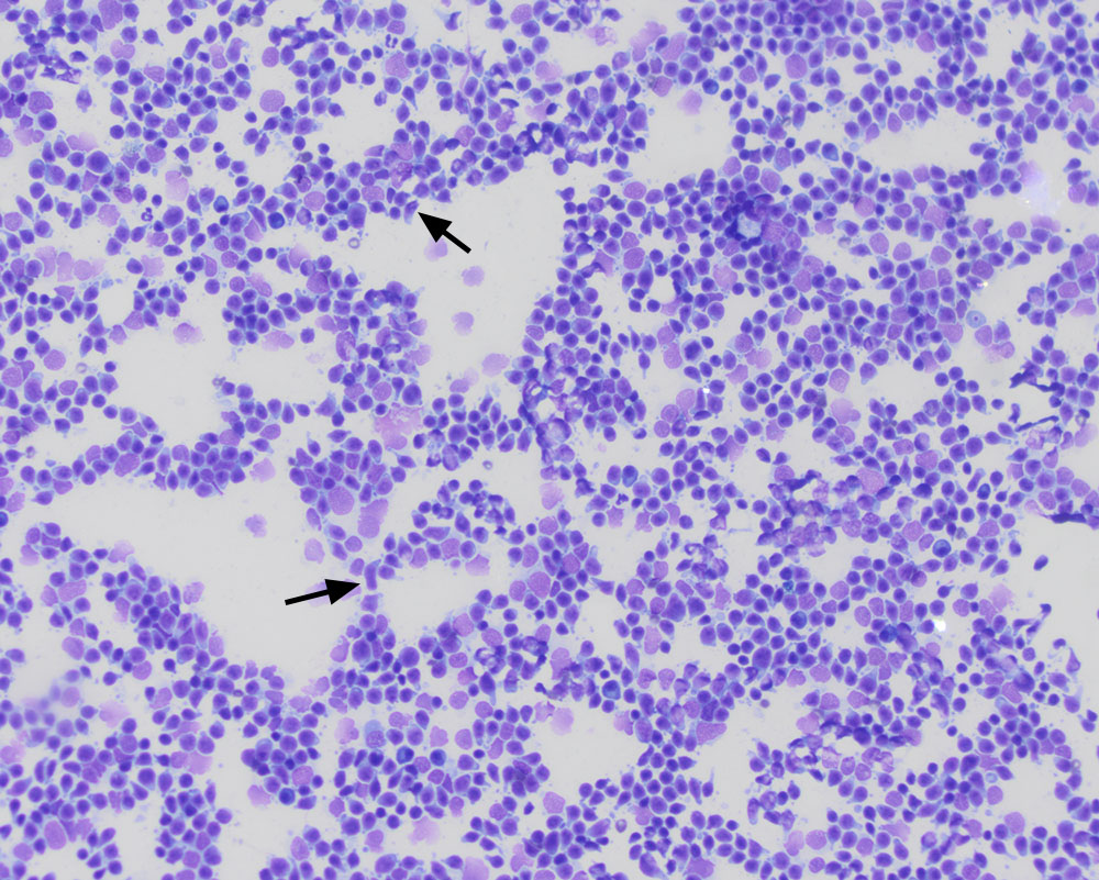
From low power, the cell population looks homogeneous, with cytoplasmic tailing (arrows), mimicking a T zone lymphoma. However, if you look carefully, you will note that the cells are intermediate to large versus small to intermediate as is typical for a T zone lymphoma (rapid stain, 20x objective)