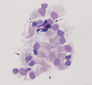
A cluster of ciliated columnar epithelial cells (arrowhead is highlighting cilia) with an interspersed goblet cell (arrow) containing numerous round purple mucin granules. There is also a lightly vacuolated macrophage (short arrow) and small lymphocyte (modified Wright’s stain). This image was from an endoscopic tracheal wash from a clinically healthy horse.

