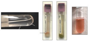
The sample on the left is of low cellularity but flecks of mucus can be seen within the fluid (white arrows). The next tube has a good sampling of mucus, which has been sedimented by centrifugation. The sample would have been cloudy grossly. It is slightly contaminated with red blood cells (black arrow) since the fluid was collected via the trachea. The third sample is green with a sediment. The green color supports oropharyngeal contamination. Although cytologic results may not be informative, freshly made smears from this tube may still reveal inflammation (although the location of the inflammation would be hard to ascertain – pharynx or lower airways). The fourth tube contains little mucus and is blood-contaminated, however such a fluid could also be yielded from a horse with hemorrhage (e.g. exercise-induced) if actively bleeding.

