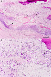
Low power photomicrographs of a representative diagnostic region of a formalin-fixed paraffin-embedded section of the excised left axillary mass from an alpaca. A: The wall of the mass transitioned from an area with prominent keratohyaline granules (infundibular differentiation, short arrow), to a region without a granular layer and amorphous keratin (isthmic differentiation, arrowhead), to epithelium with abrupt keratinization and rafts of lamellar to hyalinized keratin with ‘ghost’ cells (matrical differentiation, arrow). There was extensive disruption of the cyst wall (not shown) resulting in granulomatous inflammation with multinucleated giant cells and fibrosis (*), and multifocal osseous metaplasia (**) in reaction to the keratin. Bar = 500 μm. B: The higher magnification image illustrates the granulomatous inflammatory reaction with many multinucleated giant cells (arrows) targeting rafts of keratin with ‘ghost’ cells (arrowhead), embedded in a fibrotic stroma with osseous metaplasia (*). Bar = 50 μm. Hematoxylin & eosin (H&E) stain.

