
Indications
Indications for bone marrow sampling include:
- Evaluation of persistent or unexplained abnormalities in peripheral blood, including:
- Pancytopenia (anemia, usually non-regenerative, neutropenia, thrombocytopenia)
- Unexplained, persistent, nonregenerative anemia
- Unexplained, persistent, thrombocytopenia
- Unexplained, persistent, marked neutrophilia and/or monocytosis
- Unexplained, persistent, moderate to marked monocytosis
- Unexplained, persistent, moderate to marked eosinophilia and/or basophilia
- Unexplained, persistent, marked thrombocytosis
- Evidence of abnormal maturation with cytopenias or cytosis.
- Presence of abnormal cells in blood, e.g. blasts, mast cells, histiocytes, without a primary extramedullary mass or cause.
- Monoclonal gammopathy
- Confirmation of multiple myeloma (more in dogs and horses than cats)
- Staging of neoplasia (e.g. lymphoma, mast cell tumors)
- Evaluation for occult neoplasia or confirmation of infiltrative neoplasia, e.g. histiocytic sarcoma in dogs
- Estimation of body iron stores or to confirm iron deficiency anemia
- Evaluation for infectious organisms may be seen in bone marrow, e.g. Leishmania, Histoplasma
- Fever of unknown origin
Methods
The common sites of bone marrow collection vary with species:
- Dogs: Proximal humerus, proximal femur, iliac crest, sternum (manubrium in large dogs)
- Cats: Proximal humerus, proximal femur
- Horses and cattle: Rib, sternum (between front legs in horses), ilium
- Avian: Sternum, proximal tibiotarsus
General anesthesia or sedation with local anesthesia is required for bone marrow collection, along with pain relief. The collection must be sterile, so the site is prepared aseptically. The subcutaneous area and periosteum are infused with small amounts of local anesthetic (the periosteum can be infused by walking the needle along the bone).
Bone marrow aspirate
It is important to be prepared (get equipment and slides ready), work quickly, and make high quality smears. For an aspirate, the sample should ideally be collected directly into an anticoagulant so that it does not clot quickly and allows you time to make good quality smears. We use 1 ml of citrate, but citrate-phosphate dextrose from blood transfusion bags or 1% EDTA can be used as well. More anticoagulant can be added if the sample starts to clot. We use a 15 g and 18 g Illinios sternal-iliac needle in dogs and cats, respectively. We prefer to remove the plastic needle guard.
Procedure
- The needle is introduced into the bone by rotating until the needle is seated in the bone, then it is advanced a short distance by rotation using some pressure. The cap is removed followed by the needle stylet. The stylet should only be removed if you are in the marrow cavity.
- A 10 ml syringe containing 1 ml of anticoagulant is inserted into the needle and the plunger pulled back quickly several times. The aspirated fluid is blood mixed with multiple small bone marrow particles (a.k.a. bone marrow spicules).
- The fluid is expelled into a petri dish. The spicules sink to the surface and can be picked up using a glass pipette with a rubber bulb (in preference to a plastic pipette; spicules like to stick to the plastic). The spicules are transferred to clean glass slides (near the frosted edge) with a small amount of blood (too little or too much blood will make it difficult to spread the slides apart).
- Then a spreader slide is placed on top of the slide with the spicules and a gentle “squash” or contact smear is made. A good quality smear has a central clear area of marrow surrounded by blood (see images on right)
- Rapidly air-dry the slides (place the back of the slide on the nozzle of a hairdryer on high).
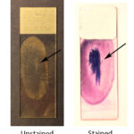
- Make as many slides as possible so additional stains can be done. We routinely stain 4 slides with Wright’s stain and 1 slide with Prussian blue for iron stores. Unstained smears can be used for other stains as needed.
- If you do not have anticoagulant, the marrow can be expelled gently onto multiple slides placed vertically (frosted edge up) resting at an angle against a stable structure. As the marrow is expelled, particles will stick to the slide and the blood will run down to the base of the slide and can be removed or blotted away. One person should expel marrow as another person quickly makes and dries the slides.
Bone marrow core biopsy
Bone marrow biopsy is performed similarly, with differences noted below, using a 13 g or 15 g Jamshidi bone marrow core biopsy in dogs and cats, respectively. These needles have a tapered end to retain the obtained core inside the bore. Studies have shown that a 13 g Jamshidi provides superior samples than a 15 g needle in dogs, whereas a biopsy is easier to perform on the humerus of cats with a 15 g needle (and produces less swelling). The humerus also provides superior samples for a core biopsy than the ilium in dogs (Abrams-Ogg et al 2012, Abrams-Ogg et al 2014).
Procedure
- Once the core biopsy needle is seated within the bone, the stylet is removed before advancing the needle deeper into the bone. You can then replace the cap.
- Advance the needle without the stylet a good distance. Slightly wiggle the end of the needle to free up the core.
- Withdraw the needle; the core will be wedged inside the needle bore. Remove the cap (if still on).
- Place the wire probe in the end of the needle and use it to push the core through the entrance of the needle (where the cap was) and onto a glass slide.
- Gently, holding one end only to reduce crush artifact, roll the core on 1-2 glass slides for cytologic evaluation. Note that cells exfoliate variably from rolled cores, with more erythroid exfoliating than myeloid precursors. This affects assessment of myeloid to erythroid ratios.
- Place the sample into formalin.
- Keep any smears used for cytologic evaluation, including blood smears, away from the core in formalin (ship separately) because formalin causes major staining artifacts.