
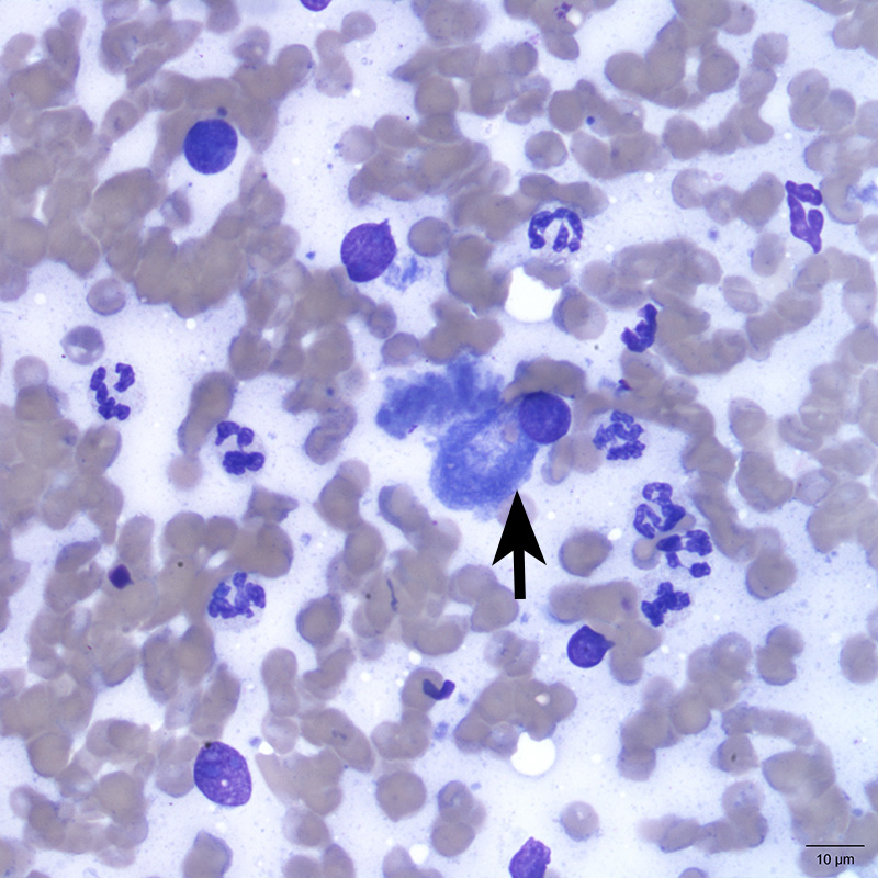
A well-differentiated osteoblast (arrow) is seen amongst non-degenerate neutrophils and few free nuclei (from ruptured cells). The neutrophils appeared convincingly increased over the expected contribution from blood supportive of inflammation. (Wright’s stain, 50x objective)