
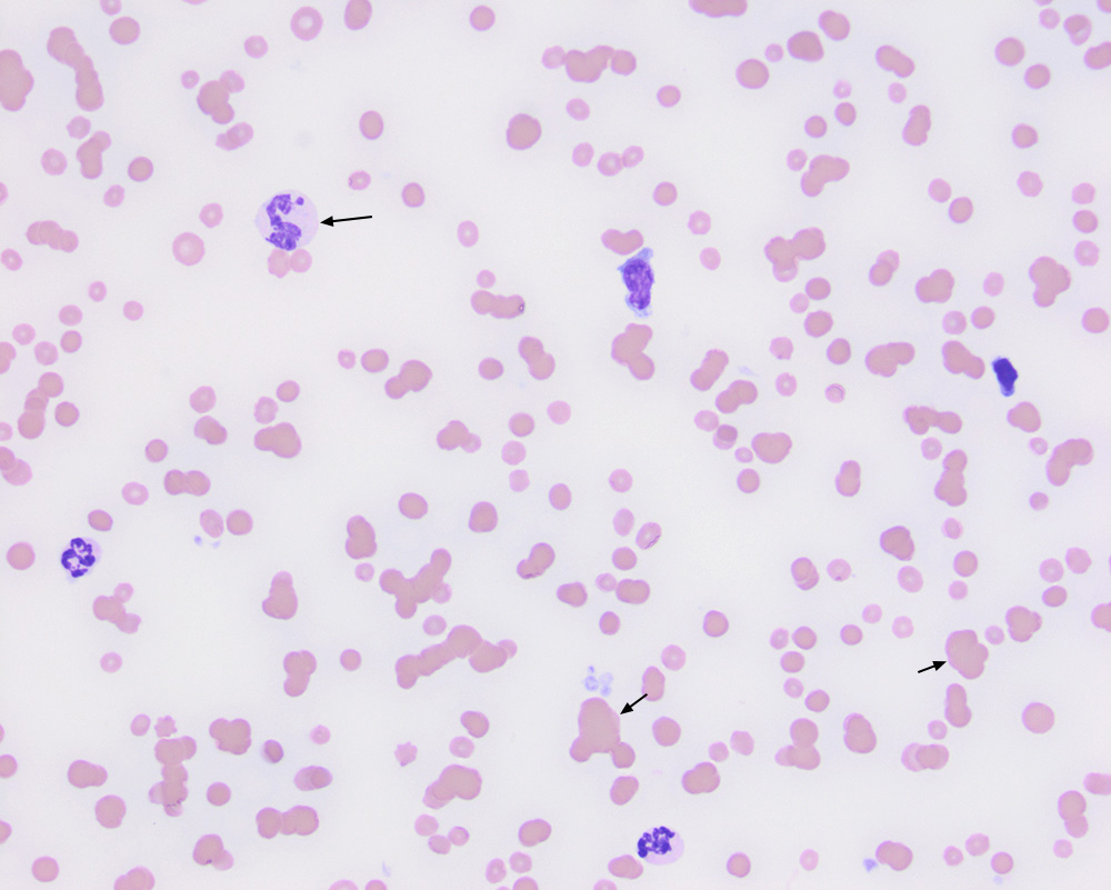
There is a giant neutrophil in this image. Three-dimensional clumps of red blood cells (agglutinates, short arrows) are present, along with short stacks (mild rouleaux formation). The other leukocytes are a monocyte (middle upper cell), lymphocyte (right upper cell), and two neutrophils (lower left and right), both of which are subtly hypersegmented (50x objective, modified Wright’s stain).