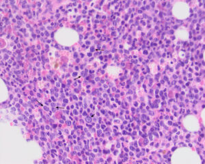
Cell identification is more difficult in a histologic section than a smear from an aspirate of bone marrow. Differentiating neutrophils and monocytes can be identified by their nuclear shapes and their cytoplasm color. Numerous blasts (large round cells with a high nuclear to cytoplasmic ratio, long arrows) are seen throughout the marrow with a few mitotic figures (short arrow). There are only low numbers of cells with small dark round nuclei and red or purple cytoplasm, corresponding to late stage erythroid progenitors (Hematoxylin & Eosin, 50x objective).

