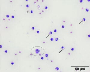
The smear consisted of many non-degenerate neutrophils (arrows) with a few macrophages (circled cell) that contained refractile material, consistent with barium, in the cytoplasm. The same material is seen free in the background (arrowhead). Modified Wright’s stain, 50x objective

