
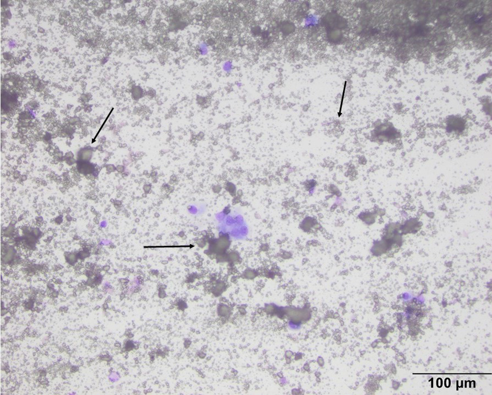
Similar mineralized material (black arrows) was seen in the smears from the aspirate of the mass over the right scapula. Note the multinucleated and mononuclear macrophages in the image (modified Wright’s stain, 20x objective). The mineral has a brownish tinge versus the pink hue in the smear from the greater trochanter mass. The pink hue in the latter is likely due to the concurrent blood contamination. Smears from such lesions often have a chalky white appearance grossly.