
Skin mass in a young dog
Case Information
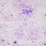
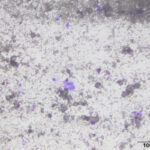
A 1-year-old male Great Dane was presented for continued monitoring of a previously diagnosed rare immune-mediated skin disease called Epidermolysis Bullosa Acquisita, which is characterized by the formation of subepidermal blisters.1 The patient was doing well and remission of the immune-mediated disease was maintained with cyclosporine and a tapering dose of methylprednisolone. During the physical examination, the patient was noted to have a firm 2 centimeter dermal mass near the right greater trochanter. A second dermal nodule, measuring about 0.75 centimeters, was noted over the right scapula approximately one month later. There was no reported discomfort noted by the owner for either mass. Cytologic evaluation of a fine-needle aspirate was performed to further investigate the origin of each mass. Stained smears from both masses had similar cytologic findings with representative images shown below.
- Given the signalment and location of the masses, what is the expected composition of the clear to light yellow-brown, coarse, elongated to amorphous material in the background?
- What differential diagnoses would you have for these cytologic findings?
|
|
|
Answers on next page