
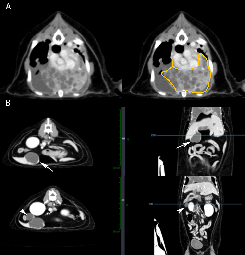
The CT scan revealed a large thoracic mass (A sagittal section, the mass is outlined by the circle in the image on the right) along with multiple pulmonary and pleural nodules (not shown). Sagittal and longitudinal sections of the abdomen showed masses associated with the liver (B, arrows, upper images) and right side of the pancreas, close to the duodenum (B, arrows, lower images).