
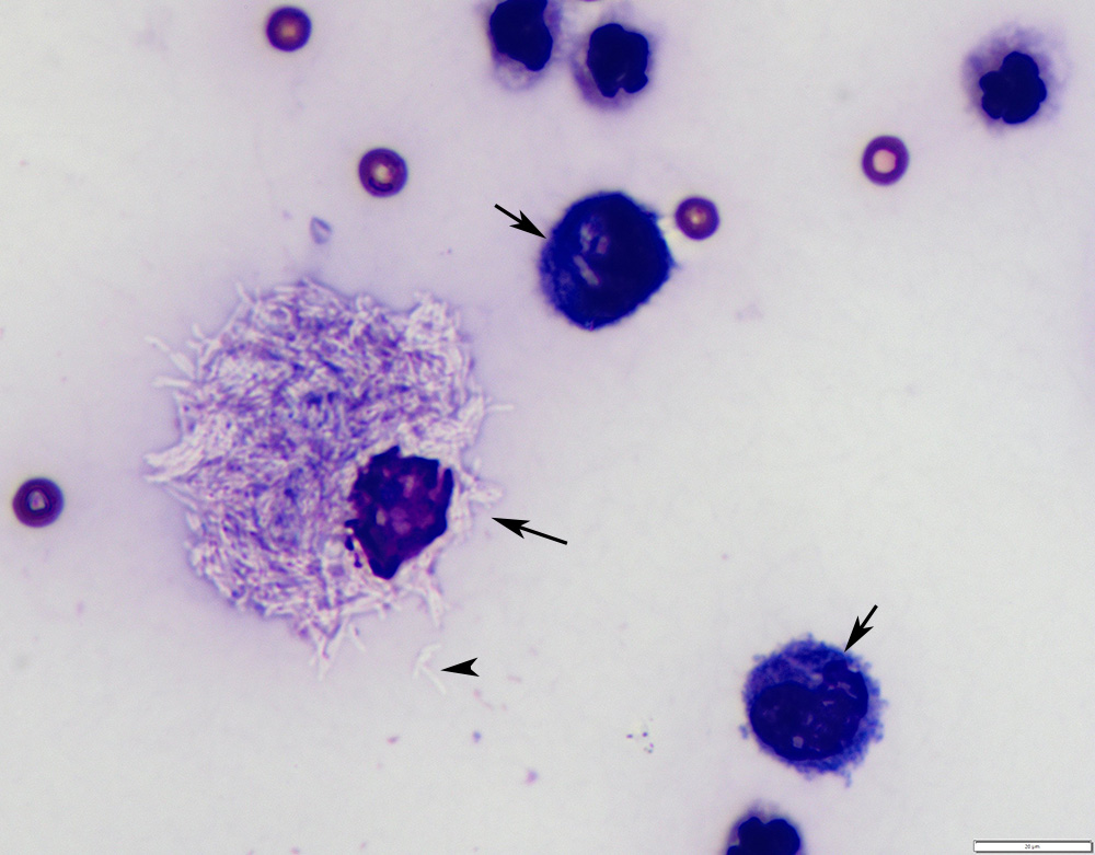
This higher power image demonstrates a macrophage that has many non-staining bacilli in the cytoplasm (long arrow). “Non-reactive” (not distended or vacuolated) macrophages also contain low numbers of similar bacilli in the cytoplasm (short arrows). Bacilli can also be seen extracellularly (from ruptured cells, arrowhead) and are refractile in appearance, which facilitates their identification (modified Wright’s stain, 100x objective)