
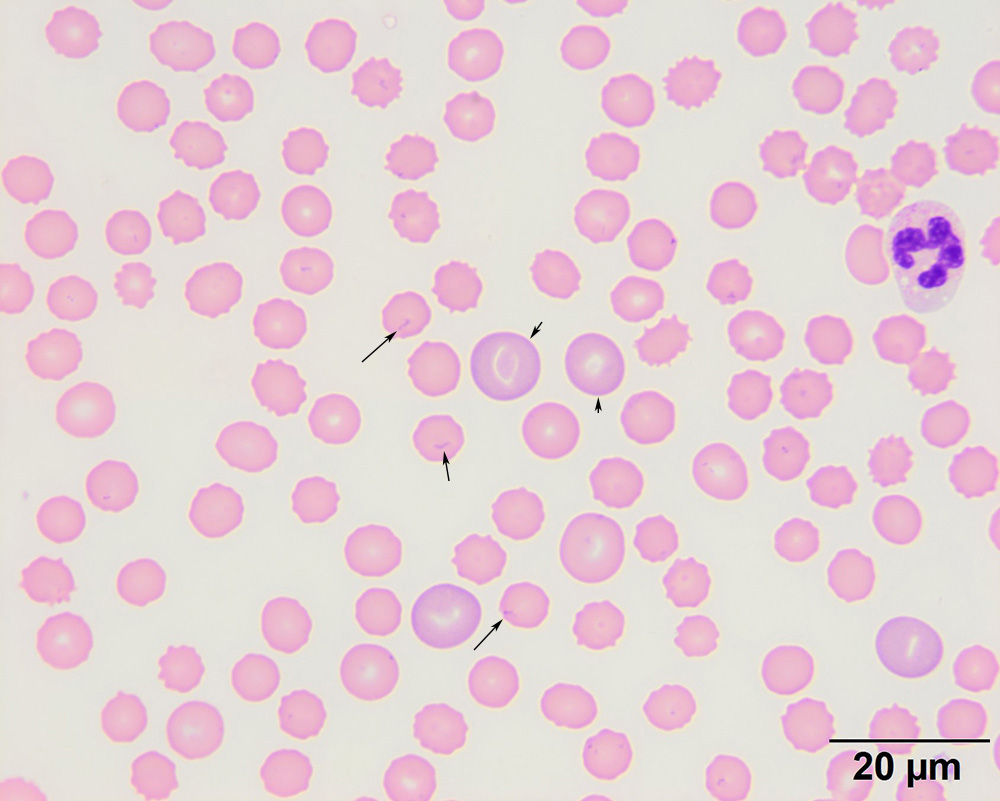
Numerous thin small organisms forming short chains or rings are seen on the surface or edges of the red blood cells (arrows). The morphologic features of the bacteria are compatible with Mycoplasma haemocanis. Note how they “pucker” or distort the red blood cell membrane, a helpful feature to differentiate them from stain precipitate or other smear artifacts. Four polychromatophils are evident in the image, supporting regeneration (two are labeled with arrowheads). There are also echinocytic red blood cells and the neutrophil has a swollen nucleus with cytoplasmic vacuoles, likely features of storage (it took a little while between diagnosis and making extra blood smears). Platelets are not present in this image, commensurate with the thrombocytopenia (modified Wright’s stain, 100x objective).