
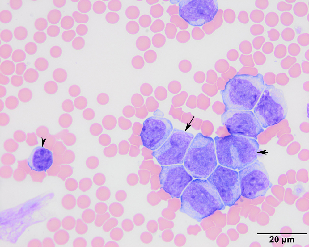
This higher power view of the tumor cells shows that low numbers have indented nuclei (long arrow) that between the rounder nuclei of blasts and more pleomorphic nuclei of mature monocytes (short arrow). Contrast the tumor cells to the small lymphocyte in the image (arrowhead) (modified Wright’s stain, 100x objective). The nuclear chromatin and cytoplasmic features of the immature cells resemble the mature monocytes (appear like variations of the same theme), leading to a presumptive diagnosis of acute myeloid leukemia.