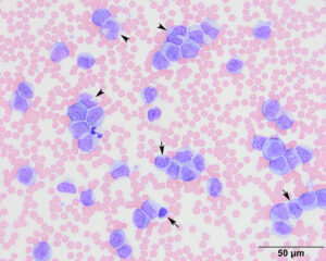
Large round cells with finely stippled chromatin and small amounts of light to medium blue cytoplasm dominated (89% of total cells), i.e. “blasts”. There were also mature monocytes (arrowheads) and cells that appeared to be differentiating towards monocytes (short arrows). There were low numbers of small mature lymphocytes, which were smaller and had darker smooth chromatin than the blasts and monocytes. Rare neutrophils were seen along with low numbers of platelets (modified Wright’s stain, 50x objective).

