
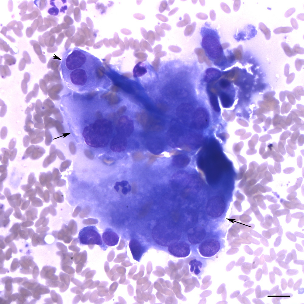
A binucleated (arrowhead) and several multinucleated giant cells (arrows) are seen in a non-cohesive grouping. There is also a spindle cell (presumptive fibroblast) on the upper right, next to the giant cell (modified Wright’s stain, scale bar = 10 um). The bi- and multi-nucleated cells display mild intracellular anisokaryosis.