
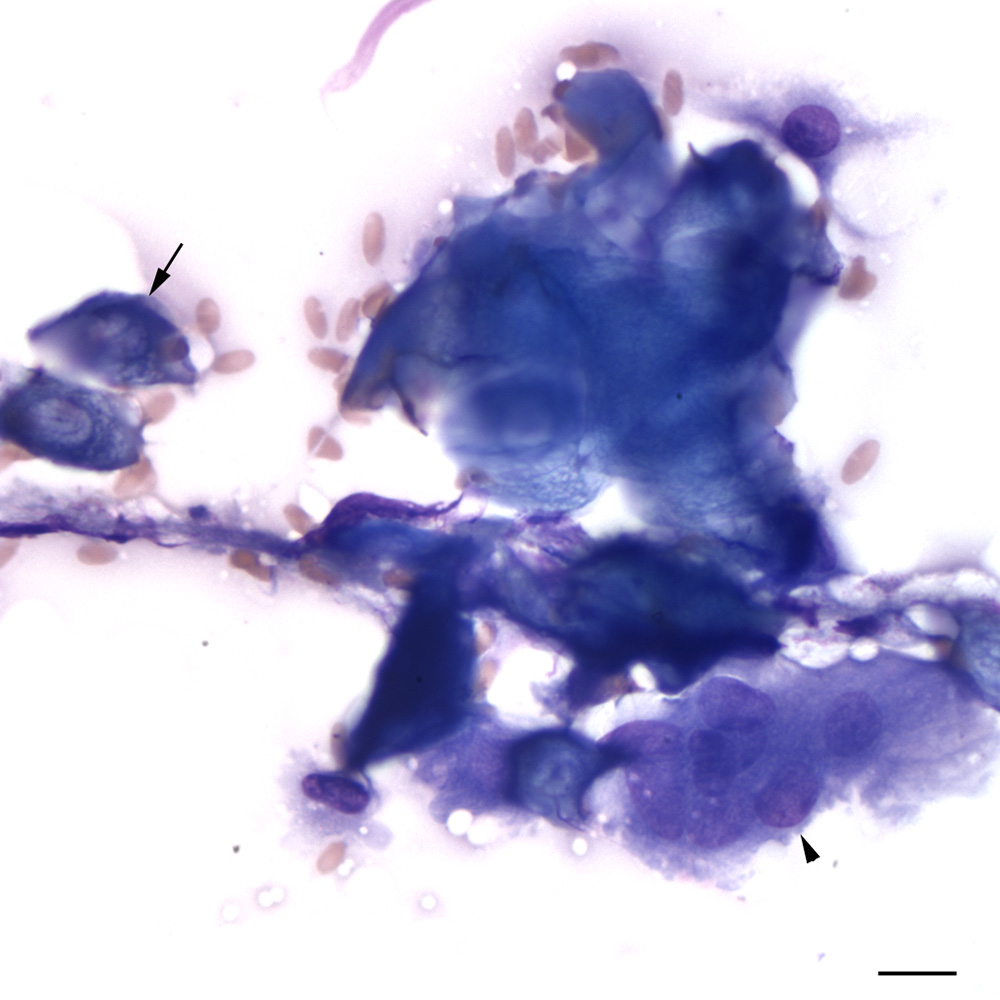
In this higher power view, you can see the central clearings corresponding to the location of a nucleus in the ‘ghost’ cells (arrow) and a multinucleated giant cell between the anucleated blue keratinized squamous cells (arrowhead) (modified Wright’s stain, scale bar = 10 um)